
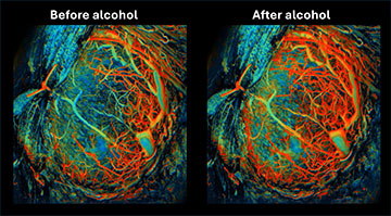
Ultrafast functional photoacoustic microscopy showed that alcohol caused a significant increase in oxygen levels in a mouse’s placenta, an effect that lasted 3 minutes. Red indicates higher levels of oxygen. [Image: Junjie Yao, Duke University]
The blood-rich placenta nourishes a growing fetus, but despite its importance, scientists still lack a full understanding of how placental problems can wreak havoc on a pregnancy―in large part due to the difficulty of capturing images of the temporary organ.
Using pregnant mice, researchers at a US university say they have developed a new approach for visualizing placental growth in vivo (Sci. Adv., doi:10.1126/sciadv.adk1278). The ultrafast imaging of the organ’s blood supply during normal and stressful conditions could shine a new light on the placenta’s crucial role in pregnancy.
Circumventing challenges
Mammalian placentas are hard to study because they are constantly moving as a patient breathes (and later in a pregnancy, as the fetus kicks), within a deep abdominal cavity. Then, once the uterus expels the placenta after birth, it no longer has a blood supply.
According to a team of scientists based at Duke University, USA, two-photon microscopy and other high-resolution optical imaging techniques aren’t able to penetrate deeply enough to study the placenta―even in mice, whose placentas have many features in common with those of humans. Furthermore, magnetic resonance imaging (MRI) and X-ray tomography don’t have the “molecular sensitivity” that would allow researchers to track changes in blood oxygenation within the placenta.
The research group, including Junjie Yao, turned to photoacoustic microscopy (PAM), which has characteristics well suited to such delicate investigation. In PAM, when a nanosecond pulsed laser beam hits tissue, the living matter absorbs the light energy and expands thermoelastically, producing a detectable ultrasonic acoustic wave that scatters less in tissue than in light waves.
The catch: the scientists found that PAM’s imaging speed is too low to resist significant blurring when the mouse subject breathes. The researchers needed 20 minutes to capture a slow-scanning placental image, resulting in severe image distortions. Thus, the team modified PAM for faster imaging rates and dubbed the result ultrafast functional PAM, or UFF-PAM.
Perfecting the techniques
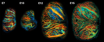
Longitudinal ultrafast functional photoacoustic microscopy images of a placenta on day 7, day 10, day 12 and day 15 of a mouse’s typically 20-day pregnancy. The diameter and density of the placental blood vessels increased by more than 200% over this period. [Image: Junjie Yao, Duke University]
The first generation of UFF-PAM employed a dual-wavelength (532 and 558 nm) pulsed laser and a ring-shaped ultrasound transducer centered at 40 MHz. Imaging elements between the two included a 12-facet polygon scanner placed after the objective lens in the signal path. However, the damping of the signal in a watery test environment introduced optical aberrations, so the team developed a second-generation UFF-PAM system with the polygon scanner placed in the air and before the objective, which resolved the issue.
The researchers also needed a window to view the placenta at different points in a mouse’s 20-day pregnancy. They surgically implanted a 10×10-mm optical window, 1 mm thick, in the living mouse’s abdominal wall. The window’s outer titanium frame clipped onto the UFF-PAM system and further reduced the distortions from the pregnant mouse’s breathing.
Scientists can remove the window from the mouse after pregnancy, according to Yao. “For human application, the concept of an ‘implanted window’ isn't directly translatable due to safety, ethical and technical challenges,” he says. “Instead, we envision noninvasive or minimally invasive imaging technologies, such as advanced ultrasound, photoacoustic or MRI techniques tailored for placental observation, which can offer insights without the need for surgical implantation.”
“One surprising finding was the crucial role of a hypoxic environment in early placental development, which protects the fetus from oxidative stress and triggers the rapid development of new blood vessels,” Yao adds. “The ability to directly observe these dynamic changes and the placenta's complex microvessel network formation in real time provided unexpected insights into the adaptive mechanisms of placental development.”
Next, the research team from Duke, joined by several other US and Chinese universities, will refine the imaging techniques for noninvasive photoacoustic imaging approaches that could be used on human patients. An example of a potential system for humans would be a transvaginal photoacoustic catheter, providing it could achieve the same level of resolution as the UFF-PAM system for mice.